Yoo, my people! Kwamé Asantè here, bubbling with excitement from Accra, where the future of medicine is not just being discussed, it is being built. Today, we are peeling back the layers on something truly monumental: the FDA-approved AI tools that are literally saving lives by detecting cancer and heart disease with incredible precision. This is not just Silicon Valley magic, my friends, this is a global phenomenon, and Ghana is right there, pushing the boundaries.
The Technical Challenge: Unveiling the Invisible Threat
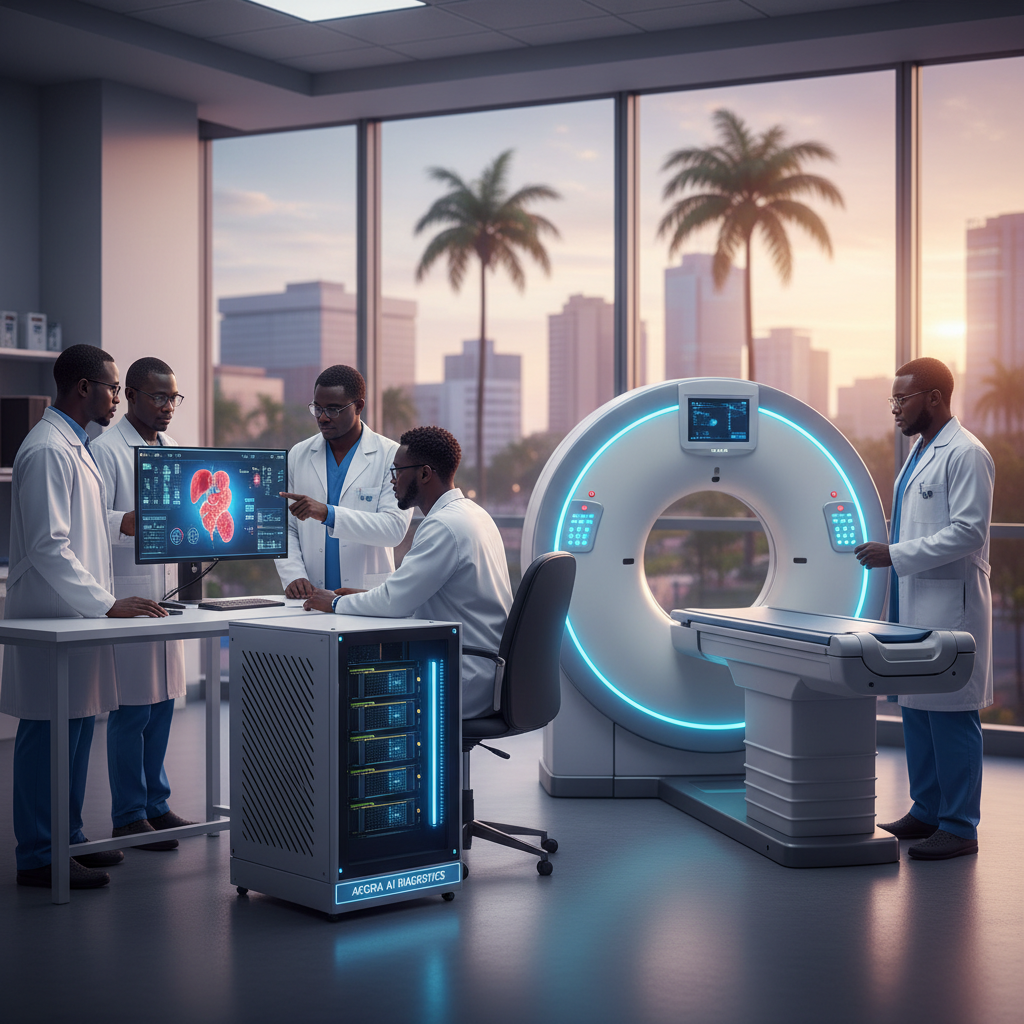
Think about the sheer volume and complexity of medical imaging data: X-rays, CT scans, MRIs, echocardiograms. Human eyes, no matter how skilled, can miss subtle anomalies, especially when fatigue sets in or patient loads are overwhelming. The problem is compounded in regions like ours, where specialist radiologists are few and far between. According to a 2023 report, sub-Saharan Africa has less than one radiologist per million people, a stark contrast to developed nations. This creates a critical bottleneck in early diagnosis, which is paramount for diseases like cancer and heart conditions. The technical challenge is clear: how do we augment human expertise, standardize diagnostic quality, and accelerate detection without compromising accuracy? The answer, my friends, is AI, specifically deep learning models trained on vast, diverse datasets.
Architecture Overview: The Diagnostic AI Pipeline
At its core, an FDA-approved AI diagnostic system is a sophisticated pipeline designed for robustness, accuracy, and interpretability. It typically comprises several key components:
- Data Ingestion and Preprocessing: Raw medical images from various modalities are anonymized, normalized, and augmented. This step is crucial for reducing noise, standardizing image intensity, and expanding the training dataset. Techniques like histogram equalization and Z-score normalization are common.
- Feature Extraction (Deep Learning): This is where the magic happens. Convolutional Neural Networks (CNNs) are the workhorses here. Architectures like ResNet, Inception, and U-Net are frequently employed. For cancer detection in pathology slides, for instance, a CNN might learn to identify cellular morphology indicative of malignancy. For cardiac MRI analysis, it could pinpoint subtle wall motion abnormalities or myocardial scarring.
- Classification/Segmentation Heads: Following feature extraction, specialized layers perform the diagnostic task. A classification head might output a probability score for 'malignant' or 'benign', while a segmentation head would delineate tumor boundaries or cardiac chambers pixel by pixel. For heart disease, this could involve segmenting the left ventricle to calculate ejection fraction, a key indicator of heart function.
- Post-processing and Clinical Integration: The AI's output is often refined with clinical rules or combined with other patient data. Crucially, these systems are designed to integrate seamlessly into existing Pacs (Picture Archiving and Communication Systems) and EMR (Electronic Medical Record) systems, providing radiologists with AI-generated insights directly within their workflow.
- Explainability (XAI) Module: Given the critical nature of medical diagnosis, 'black box' models are unacceptable. Many FDA-approved systems incorporate XAI techniques like Grad-CAM or Lime to highlight the specific regions in an image that influenced the AI's decision, fostering trust and aiding clinician review.
Key Algorithms and Approaches: The Brains Behind the Breakthroughs
For cancer detection, especially in radiology, object detection models like Faster R-cnn or Yolo (You Only Look Once) are adapted to identify suspicious lesions. Imagine a model scanning a mammogram and drawing bounding boxes around potential tumors, flagging them for human review. For subtle changes, segmentation networks like U-Net are invaluable. These pixel-level predictions are critical for quantifying tumor size or identifying early-stage disease.
For heart disease, particularly in echocardiography or cardiac MRI, recurrent neural networks (RNNs) or 3D CNNs are sometimes used to analyze sequences of images, capturing the dynamic nature of the beating heart. This allows for the assessment of cardiac function over time, detecting issues like weakened heart muscle or valve dysfunction. Transfer learning is also paramount; models pre-trained on massive natural image datasets (like ImageNet) are fine-tuned on medical images, leveraging their learned feature hierarchies.
Let us consider a simplified conceptual example for lung nodule detection:
def detect_lung_nodules(ct_scan_image):
 # 1. Preprocessing: Normalize intensity, resize
 processed_image = preprocess(ct_scan_image)

# 2. Feature Extraction: CNN backbone (e.g., ResNet-50)
 features = cnn_model.extract_features(processed_image)

# 3. Region Proposal Network (RPN): Propose potential nodule locations
 regions_of_interest = rpn.propose_regions(features)

# 4. Classification & Refinement: For each region, classify and refine bounding box
 final_detections = []
 for roi in regions_of_interest:
 class_label, confidence_score, bbox = detection_head.predict(roi, features)
 if class_label == 'nodule' and confidence_score > threshold:
 final_detections.append({'location': bbox, 'likelihood': confidence_score})

# 5. Explainability: Generate heatmap of influential pixels
 explanation_map = grad_cam.generate(processed_image, final_detections)

return final_detections, explanation_map
Implementation Considerations: From Lab to Life-Saving Tool
Building these systems is not for the faint of heart. Data curation is a monumental task, requiring expert annotation. Ethical considerations around data privacy, bias, and algorithmic fairness are paramount. Performance is also key; these models often demand significant computational resources. This is where NVIDIA's GPUs, like the A100 and H100, become indispensable. Training these deep learning models on terabytes of medical images would be impractical without their parallel processing power. Deployment requires robust MLOps practices, continuous monitoring, and strict version control to maintain FDA compliance.
One major trade-off is between sensitivity and specificity. A highly sensitive model might catch every potential anomaly but generate many false positives, leading to unnecessary follow-up procedures. A highly specific model might reduce false positives but risk missing true disease. Balancing these metrics is a critical part of the development and validation process.
Benchmarks and Comparisons: Raising the Bar
How do these AI systems stack up? Phenomenally! For example, an FDA-approved AI for diabetic retinopathy detection has demonstrated sensitivity and specificity comparable to, and in some cases exceeding, human ophthalmologists. For breast cancer detection in mammography, AI has shown up to a 10% improvement in detection rates compared to human readers alone, reducing false negatives. The numbers don't lie, these tools are making a tangible difference.
Traditional CAD (Computer-Aided Detection) systems, which relied on hand-crafted features and simpler machine learning, often suffered from high false positive rates. Deep learning, with its ability to learn hierarchical features directly from raw data, has fundamentally surpassed these older approaches, offering superior accuracy and generalizability.
Code-Level Insights: The Toolkit for Tomorrow's Doctors
Developers working on these systems often leverage frameworks like TensorFlow and PyTorch for model development. Libraries such as Pydicom are essential for handling Dicom medical image formats. For deployment, containerization technologies like Docker and orchestration tools like Kubernetes are common, ensuring scalability and reproducibility. NVIDIA's Monai (Medical Open Network for AI) framework is gaining significant traction, providing domain-specific tools and pre-trained models for medical imaging. It is an absolute game-changer for accelerating research and development in this space. You can find out more about these innovations on TechCrunch.
Real-World Use Cases: Impacting Lives Now
- Qure.ai's qXR for Chest X-rays: This FDA-cleared AI detects abnormalities like tuberculosis, pneumonia, and lung cancer on chest X-rays. It is deployed in over 75 countries, including several in Africa, providing rapid screening in high-volume settings. Imagine the impact in rural Ghanaian clinics where radiologists are scarce; a quick AI scan can triage urgent cases.
- Aidoc's AI for Acute Conditions: Aidoc offers a suite of FDA-cleared AI solutions that flag critical findings on CT scans, such as intracranial hemorrhage, pulmonary embolism, and cervical spine fractures. By prioritizing these urgent cases, it significantly reduces the time to diagnosis and treatment, potentially saving lives.
- HeartFlow Ffrct: This AI-powered software creates a 3D model of a patient's coronary arteries from a standard CT scan and simulates blood flow to assess the functional impact of blockages. It is FDA-approved and helps clinicians determine if a patient needs an invasive procedure, reducing unnecessary interventions.
- Google Health's Dermatology Assist: While not strictly cancer or heart disease, Google's AI for dermatology, which can identify skin conditions, including potential skin cancers, demonstrates the breadth of AI's diagnostic capabilities. This is bigger than anyone realizes, extending specialized care to underserved communities.
Gotchas and Pitfalls: Navigating the Complexities
Despite the incredible promise, challenges remain. Data bias is a serious concern; if training data does not adequately represent diverse populations, the AI may perform poorly on underrepresented groups. This is particularly relevant for Africa, where unique genetic and environmental factors can influence disease presentation. Regulatory hurdles are also significant, with the FDA requiring rigorous validation for each AI model. Maintaining model performance over time, especially as medical imaging equipment evolves, requires continuous monitoring and retraining. Furthermore, the ethical implications of AI in diagnosis, including accountability and informed consent, demand ongoing discussion and careful policy development. For deeper analysis on these ethical considerations, MIT Technology Review often publishes excellent articles.
Resources for Going Deeper: Your Journey into Medical AI
For those eager to dive further, I recommend exploring the Monai framework documentation, reading research papers on arXiv, particularly in the cs.CV and eess.IV categories for computer vision and image processing applications in medicine, and following the work of leading AI in healthcare companies. The annual Radiological Society of North America (rsna) conference is a fantastic source for the latest advancements. You can also explore academic courses on medical imaging AI offered by institutions like Stanford or MIT. The future is bright, and the opportunities for innovation, especially here in Ghana, are limitless. Let us build it together! This is our time to shine, to leverage technology not just for profit, but for profound human impact.